Monday, May 4, 2020

PROMOTING HYGIENE TO MINIMIZE THE INFECTIONS


Promoting Hygiene To Minimize the Infections  

                            University of Sheffield Researchers Discover New Compound Kills ...

Introduction

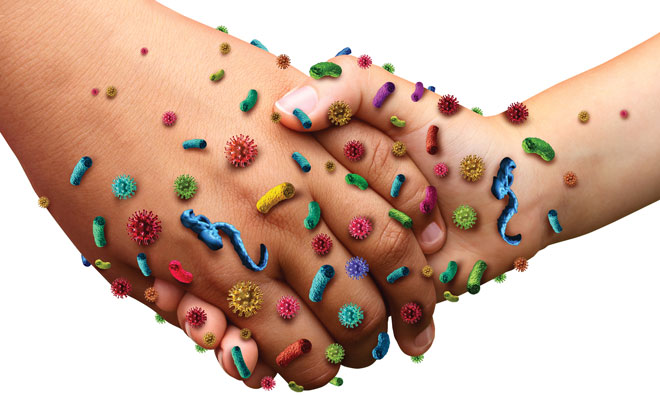
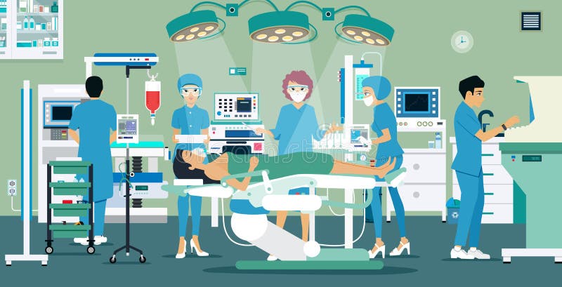
Healthcare Associated Infections (HCAI's) can occur following direct contact within healthcare settings or following surgery. These infections, the majority of which are preventable, can cause significant harm to those affected, increase hospital stay and put added pressure on the NHS.

There are many reasons a patient may develop a Health care associated infections, including weakened immune deference due to treatment for cancer or leukemia, complex surgical operations, insertion of medical devices such as catheters, or the over-use of antibiotics. The latter can result in the development of resistance such as meticillin-resistant Staphylococcus aureus (MRSA) making treatment with common antibiotics difficult. The increase in patients with chronic illnesses, such as diabetes, heart and kidney problems, and the increase in the elderly population are also contributing factors that predispose patients to infections.

Infection prevention and control measures, such as appropriate hand hygiene, environmental cleanliness, basic precautions during invasive procedures, and education of staff, patients and visitors, are simple and inexpensive measures to help reduce the risk of HCAI's.


How infections are spread/Mode of transmission

When healthcare staff are educated about the ways infections are spread it helps them to decide what preventative measures they need to take and they are better placed to inform patients. Spread of infection requires three elements. The first is an infectious agent - a bacteria, virus, fungi or protozoa. The source may be the patients’ own flora, other patients, staff or visitors, or contaminated equipment. The second element is a susceptible host –someone prone to infections because of an underlying disease, surgical procedures or indwelling devices, and the final factor is a means of transmission.

                      Cleanliness of health care facilities - Basics — Vikaspedia

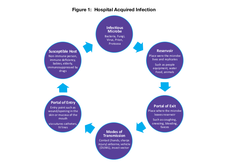
There are four main routes of infection transmission:

Contact transmission (direct or indirect):

Direct: body surface to body surface contact and physical transfer of microorganism between an infected or colonized (carrier) person to another or from one site to another in the same individual.

Indirect: contact between a person and contaminated surface or object.

Airborne /droplet transmission:

Droplet transmission: large droplets produced during coughing, sneezing, talking and suctioning generate droplets that land on surfaces which when touched can contaminate hands.

Airborne transmission: smaller micro-organisms, contaminated water particles or airborne dust particles containing the infectious agent are dispersed by air currents then inhaled or deposited onto horizontal surfaces, equipment etc.

Food and water (Fecal–oral) transmission:

Organisms can be transmitted via the food we eat and handle e.g. Campylobacter on raw chicken, by inappropriate handling of contaminated raw food or inadequate cooking. Cross-infection can occur via contaminated surfaces or infected food handlers if their hands are not cleaned after using the toilet. Water provides an ideal breeding ground for some micro- organisms, which can then be ingested, or as in Legionella inhaled.

Vector borne transmission:

These are infections transmitted by, flies, mosquitoes, and rats.
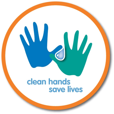
Hand hygiene

Hand hygiene is the single most important measure in reducing the risk of transmission of micro-organisms from one person to another or from one site to another on the same person. The World Health Organisation advises that cleaning hands promptly and as thoroughly as possible between patient contacts and after contact with blood, body fluids, secretions, excretions and contaminated equipment, is essential in order to minimize the risk of cross-infection in healthcare.

                        SG Infection Control (@StGInfectionCon) | Twitter
The Infection Prevention Society advises that effective hand hygiene involves the use of soap and water or alcohol hand rub. Liquid soap and warm running water can be used to mechanically remove transient micro-organisms from the hands and is perfectly acceptable for the vast majority of healthcare interventions. An alcohol-based product can be used for general hand hygiene in place of a hand-wash but only if hands are visibly clean. Alcohol rub, soap, water and paper towels must be available to encourage hand hygiene, and sinks for hand washing should be used for only that and not washing tea cups, dentures etc. A poster demonstrating the correct hand wash/rub techniques should be displayed at each clinical hand wash basin and a poster encouraging hand washing should be displayed in all patient and visitor toilets.

When building or refurbishing any health care environment provision of proper hand washing facilities must be a priority, therefore the Infection Prevention and Control Nurse (IPCN) should be involved at planning stage.

Environmental cleanliness in healthcare

Contaminated healthcare surfaces play a huge role in the transmission of dangerous pathogens, including Clostridium difficile and MRSA. 

                        Environmental Services - APIC


These pathogens are capable of prolonged survival on surfaces, therefore appropriate cleaning of those surfaces and equipment which patients and healthcare staff touch, is necessary to reduce transmission. Frequently touched surfaces, such as taps, door handles and light switches are more likely to harbor and transmit micro-organisms, so it’s important to ensure these surfaces are cleaned frequently in line with specifications for cleanliness in the NHS.

Public perception is very important and if the environment in which patients or their relatives are cared for appears unclean, it can lead to the perception that care is equally bad.

What patients and visitors can do to help in preventing HCAI's
While it is commonly perceived that HCAI's are attributable to healthcare staff, it is worth remembering that patients and their visitors also contribute. There are some simple things that patients and visitors should be encouraged to do in the fight to prevent transmission of infections.

Patients can: wash hands after using the toilet and before meals; cover mouth and nose with tissue when sneezing or coughing, bin the tissue and wash hands; if socializing at another patient’s bed space, clean hands afterwards; ask staff if they have washed their hands and/or changed their gloves before doing your care; wash/shower daily using own toiletries; keep locker and table top tidy and free from clutter to help with cleaning; do not interfere with drips, drains or dressings; and report any concerns about cleanliness or care standard.

Additionally, visitors can: use the hand sanitizer when entering and leaving the hospital ward or department; not visit if you are unwell especially with colds/flu or stomach upsets; avoid bringing very young children to visit; no not bring in fresh flowers or plants; speak to staff before bringing food into the hospital; and report any concerns about cleanliness or patient care.

It is only by staff, patients and visitors working together to do their bit that we will win the fight to reduce HCAI's.


WHO Recommendations: -

                        WHO | Guidelines on core components of infection prevention and ...


Conclusion: - Good Hygiene Practices Reducing the Spread of Infections and Viruses. The most important way to reduce the spread of infections is hand washing - always wash regularly with soap Providing boxes of tissues and encourage their use.


                        Infection Prevention is Everyone's Business...That means YOU ...

Written By : -
Priyanka Massey 
Certified Infection Control Nurse,
Professional Trainer
Ingenious Health Care Consultants Pvt.Ltd.

Thanks for the attention.
Reference taken from Wikipedia


Friday, May 1, 2020

Operation Theater


Operation Theater 

                          Operating Room stock vector. Illustration of surgical - 82728773

Introduction

An operating theater is a facility within a hospital where surgical operations are carried out in an aseptic environment. Historically, the term "Operating Theater" referred to a non-sterile, tiered theater or amphitheater in which students and other spectators could watch surgeons perform surgery. 

Operation Theater is a facility within a hospital where surgical operations are carried out in a sterile environment.The Operation Theater department pride themselves on delivering high quality of care in a clean and friendly environment, delivered by teams with a vast range of expertise, for each specialty.

An operation theater complex is the "heart" of any major surgical hospital. An operating theater, operating room, surgery suite or a surgery center is a room within a hospital within which surgical and other operations are carried out. Operating theater were so-called in the United Kingdom because they traditionally consisted of semi-circular amphitheaters to allow students to observe the medical procedures .

The patient is the center point of a functioning OT complex. He / she is in isolation for varying times, away from his near and dear ones and is physically sick. Efforts are directed to maintain vital functions, prevent infections / promote healing with safety, comfort and economy.

A room in hospital which contains an operating table or similar device, generally accommodating one patient at a time, during which, under the direct supervision of a medical or dental practitioner, the patient undergoes an operative procedure for the prevention, cure, relief or diagnosis of disease, or in pregnancy, childbirth. An operating theater must: allow positioning of the patient on the table or device so as to render the operation possible or convenient; have adjustable lighting powerful enough to permit fine or delicate work under aseptic conditions; provide sterile instruments and facilities for staff to change clothing; provide deeper pain relief than the basic sedation administered in the ward or local anesthetic.

The establishment and working of the Operation Theater (O.T.) needs specialized planning and execution and is not a simple civil engineering work. A "civil-mechanical-electrical-electronic- bio medical" combo effort driven and coordinated by the needs, preferences and safety of the medical/ surgical team forms the basis for starting and maintaining an operation theater. Anesthesiologists, by virtue of their knowledge of the intricacies of physiology, physics and biomedical aspects of medicine and constant proximity to the operation theater should preferably be involved from the early stages of planning of operating theaters.

144 Anaesthesia Stock Illustrations, Cliparts And Royalty Free ...

Purpose of Operation Theater Complex


OT complexes are designed and built to carry out investigative, diagnostic, therapeutic and palliative procedures of varying degrees of incisiveness. Many such set ups are customized to the requirements based on size of hospital, patient turnover and may be specialty specific. The aim is to provide the maximum benefit for maximum number of patients arriving to the operation theater.. Both the present as well as future needs should be kept in mind while planning.

Different zones of O.T Complex 

The location and flow of the patients, the staff and the materials form the three broad groups to be considered during all stages of design.

Four zones can be described in an O T complex, based on varying degrees of cleanliness, in which the bacteriological count progressively diminishes from the outer to the inner zones (operating area) and is maintained by a differential decreasing positive pressure ventilation gradient from the inner zone to the outer zone.

(1) Protective zone: It includes

Change rooms for all medical and paramedical staff with conveniences.
Transfer bay for patient, material & equipment's.
Rooms for administrative staff.
Stores & records.
Pre & post-operative rooms.
I.C.U. and P.A.C.U.
Sterile stores.

(2) Clean zone : Connects protective zone to aseptic zone and has other areas also like

Stores & cleaner room.
Equipment store room.
Maintenance workshop.
Kitchenette (pantry).
Firefighting device room.
Emergency exits.
Service room for staff.
Close circuit TV control area.

(3) Aseptic zone - Includes operation rooms (sterile)

(4) Disposal zone - Disposal areas from each OR & corridor lead to disposal zone.

Medical staff in operation theater Vector Image - 1521055 ...


   Sub areas (excluding OT place)

(1) Pre-operative check in area (reception)- This is important with respect to maintaining privacy, for changing from street clothes to gown and to provide lockers and lavatories for staff.

(2) Holding area- This area is planned for IV line insertion, preparation, catheter / gastric tube insertion, connection of monitors, & shall have O 2 and suction lines. Facility for CPR should be available in this area.

(3) Induction room - (anesthetic room). It should have all facilities as in OT, but there is controversy as to its need. One for each OT is required, ideally each is a duplicate of the other in each floor.

The anesthetic room will provide a more tranquil atmosphere to the patient than the OT. It should provide space for anesthetic trolleys and equipment and should be located with direct access to circulation corridors and ready access to the operating room. It will also allow cleaning, testing and storing of anesthesia equipment. It should contain work benches, sink(s). It should have sufficient power outlets and medical gas panels for testing of equipment.

(4) Post anesthetic care units (PACU) - preferably adjacent to recovery room. These should contain a medication station, hand washing station, nurse station, storage space for stretchers, supplies and monitors / equipment and gas, suction outlets and ventilator. Additionally 80 sq ft (7.43 sq m) for each patient bed, clearance of 5 ft (1.5 m) between beds and 4 ft (1.22m) between patient bed sides and adjacent walls should be planned.

(5) Staff room - Medical staff Men and women change dress from street cloth to OT attire; lockers and lavatory are essential; rest room TV, etc. are desirable.

(6) Sanitary facility for staff- One wash basin and one western closet (WC) should be provided for 8-10 persons. Showers and their number is a matter of local decision. Inclusion of toilet facilities in changing rooms is not acceptable; they should be located in an adjacent space .

(7) The anesthesia gas / cylinder manifold room / storage area- A definite area to be designated. It should be in a cool, clean room that is constructed of fire resistant materials. Conductive flooring must be present but is not required if non inflammable gases are stored. Adequate ventilation to allow leaking gases to escape, safety labels and separate places for empty and full cylinders to be allocated.

(8) Offices - for staff nurse and anesthesia staff- The office should allow access to both unrestricted and semi-restricted areas as frequent communication with public is needed.

(9) Rest rooms- Pleasant and quiet rest for staff should be arranged either as one large room for all grades of staff or as separate rooms both have merits. Comfortable chairs, one writing table, a book case etc., may be arranged.

(10) Laboratory - Small lab. with refrigerator for pathologist to be arranged.

(11) Seminar room- Since staff cannot leave an OT complex easily, it is better to have a seminar room within the OT complex. Intra-departmental discussions, teaching and training sessions for staff (with audio-visual aids) may be conducted here.

(12) Store room- This is designed to store large but less frequently used equipment in the OT. There should be storage space for special equipment after cleaning.

(13) Theater sterile supply unit (TSSU)- Within this area, following are desirable : -

Temperature between 18 0 -22 0 C, humidity of 40%­-50% is the aim.
Air conditioned with 10-12 air exchanges per hour
Storage of sterile drapes, sponges, gloves, gowns and other items ready to use.
Option to store in from one side and remove from other side.
Proper inventory to prevent running out of stock.

(14) Scrub room- This is planned to be built within the restricted area. Elbow operated or infrared sensor operated taps / water source is ideal. It is essential to have non slippery flooring in this area.

Types of O.T Complex

There are three main categories of operating the areas 

The single theater suite with OT, scrub-up and gowning, anesthesia room, trolley preparation, utility and exit bay plus staff change and limited ancillary accommodation.

The twin Theater suite with facilities similar to 1(one), but with duplicated ancillary accommodation immediate to each OT, sometimes sharing a small post anesthesia recovery area.

OT complexes of three or more OT's. with ancillary accommodation including post anesthesia recovery, reception, porter's desk, sterile store and staff change.

                         Medical surgery concept cartoon hospital Vector Image


Operating Room Equipment's 

The operating table in the center of the room can be raised, lowered, and tilted in any direction.

The operating room lights are over the table to provide bright light, without shadows, during surgery.

The anesthesia machine is at the head of the operating table. This machine has tubes that connect to the patient to assist them in breathing during surgery, and built-in monitors that help control the mixture of gases in the breathing circuit.

The anesthesia cart is next to the anesthesia machine, It contains the medications, equipment, and other supplies that the anesthesiologist may need.

Sterile instruments to be used during surgery are arranged on a stainless steel table.

An electronic monitor (which records the heart rate and respiratory rate by adhesive patches that are placed on the patient's chest).

The pulse-oximeter machine attaches to the patient's finger with an elastic band aid. It measures the amount of oxygen contained in the blood.

Automated blood pressure measuring machine that automatically inflates the blood pressure cuff on patient's arm.


An electrocute machine uses high frequency electrical signals to cauterize or seal off blood vessels and may also be used to cut through tissue with a minimal amount of bleeding.


Surgeon and assistants' equipment's 

People in the operating room wear P.P.E (personal protective equipment) to help prevent bacteria from infecting the surgical incision. This P.P.E includes the following:

Similar to normal clean-rooms, germ-less.

A protective cap covering their hair.

Masks over their lower face, covering their mouths and noses with minimal gaps to prevent inhalation of plume or airborne microbes
Shades or glasses over their eyes, including specialized colored glasses for use with different lasers. a fiber-optic headlight may be attached for greater visibility.

Sterile gloves; usually latex-free due to latex sensitivity which affects some health care workers and patients.

Long gowns, with the bottom of the gown no closer than six inches to the ground.

Protective covers on their shoes.

If x-rays are expected to be used, lead aprons/neck covers are used to prevent overexposure to radiation.


The surgeon may also wear special glasses that help him/her to see more clearly. The circulating nurse and anesthesiologist will not wear a gown in the OR because they are not a part of the sterile team. They must keep a distance of 12-16 inches from any sterile object, person, or field.

                             Comment: Tackling malware in medical equipment | E&T Magazine


Written By:
Priyanka Massey
Certified Infection Control Nurse & Professional Trainer
Ingenious Health Care Consultants Pvt.Ltd.

Reference taken from Wikipedia
Thanks for Your attention. 

Thursday, April 30, 2020

BIO MEDICAL WASTE MANAGEMENT


BIO-MEDICAL WASTE MANAGEMENT


Introduction

Biomedical waste (BMW) is any waste produced during the diagnosis, treatment, or immunization of human or animal research activities pertaining thereto or in the production or testing of biological or in health camps. It follows the cradle to grave approach which is characterization, quantification, segregation, storage, transport, and treatment of BMW.

The basic principle of good BMW practice is based on the concept of 3Rs, namely, reduce, recycle, and reuse. The best BMW management methods aim at avoiding generation of waste or recovering as much as waste as possible, rather than disposing. Therefore, the various methods of BMW disposal, according to their desirability, are prevent, reduce, reuse, recycle, recover, treat, and lastly dispose. Hence, the waste should be tackled at source rather than “end of pipe approach.”

BMW treatment and disposal facility means any facility wherein treatment, disposal of BMW or processes incidental to such treatment and disposal is carried out.

Only about 10%–25% of BMW is hazardous, and the remaining 75%–95% is nonhazardous. The hazardous part of the waste presents physical, chemical, and/or microbiological risk to the general population and health-care workers associated with handling, treatment, and disposal of waste.

In a World Health Organization (WHO) meeting in Geneva, in June 2007, core principles for achieving safe and sustainable management of health-care waste were developed. It was stressed that through right investment of resources and complete commitment, the harmful effects of health-care waste to the people and environment can be reduced. All stakeholders associated with financing and supporting health-care activities are morally and legally obliged to ensure the safety of others and therefore should share in the cost of proper management of BMW. In addition, it is the duty of manufacturer to produce environment-friendly medical devices to ensure its safe disposal. WHO reinforced that government should designate a part of the budget for creation, support, and maintenance of efficient health-care waste management system. These include novel and ingenious methods/devices to reduce the bulk and toxicity of health-care waste. Nongovernmental Organization should undertake program and activities that contribute in this incentive.

Amendments in BMW rules

The first edition of WHO handbook on safe management of wastes from health-care activities known as “The Blue Book” came out in 1999. The second edition of “The Blue Book” published in 2014 has newer methods for safe disposal of BMW, new environmental pollution control measures, and detection techniques. In addition, new topics such as health-care waste management in emergencies, emerging pandemics, drug-resistant bacteria, and climate changes were covered in the second edition.

                               BMW Rules 2016 (Amendments) | medCampus

The new biomedical waste management rules have been notified to efficiently manage BMW in the country in 2016 These rules have been modified to include the word handling and bring more clarity in the application. In addition, strict rules have been made to ensure no pilferage of recyclables item, no secondary handling or in advent scattering or spillage by animals during transport from the Health care facility's to the common BMW treatment facility. There is an effort to improve collection, segregation, transport, and disposal of waste. Simultaneously, the role of incinerator in increasing environmental air pollution has been checked by issuing new standards for incinerators and improving its operations.


Benefits of the new biomedical waste rules as per 2016 guidelines 

The new rules are stringent and elaborate and should bring about a change in the way, the BMW is being managed in India. Under the new rules, coverage has increased to include various health-care related camps such as vaccination camps, blood donation camps, and surgical camps.
                                            
                            What are the benefits of waste management? - Quora

Another distinction is in the segregation, packaging, transport, and storage of BMW waste. The categories have been reduced to four to bring about ease of segregation. One of the main principle of disposal of BMW is that segregation has to be done at the source of generation of the waste. To overcome confusion created by large number of categories, this has been simplified to make it convenient and manageable for all HCWs. Now, the color coding (i.e., yellow, red, white, and blue) of the bags/containers is linked to a particular type of waste and its specific treatment option. For example, the disposal of chemical solid waste and cytotoxic waste to be done in yellow bag which goes for incineration/plasma pyrolysis/deep burial.

In addition, the health care fascility has to do pre-treatment of various laboratory waste and blood bags according to guidelines of WHO and NACO, to decrease chances of infections being transmitted to HCWs handling waste at treatment stage.
Within 2 years, plastic bags, gloves, and blood bags have to be phased out to eliminate emissions of dioxins and fur-ans during their burning into the environment. The new rule also calls for a bar code system for all bags/containers used for BMW treatment and disposal. This step will help in tracking and identifying bags during inspection for quality control and also quality assurance.

The BMW in red/blue bag or container which is for recycling will be sent only to an authorized recycler. This will keep the recycler in realm and in control of various government agencies. Greater emphasis has been given to recycling of waste to conserve resources as well as decrease pollution.

The 2016 guidelines are more specific regarding the dependence of health care facility on common biomedical waste treatment facility will provide land for setting up Common bio medical treatment facility State government or UT government will provide land for setting up Common bio-medical waste treatment facility. and no occupier of an Health care facility shall establish an on-site treatment and disposal facility if a Common bio-medical waste treatment facility is available within 75 kms. 

This has several advantages as installation and functioning of individual BMW treatment facility as well as recruiting separate, dedicate, and skilled workforce require high capital investment. Common bio-medical waste treatment facility is a popular concept in developed countries because by operating it at its full potential, the cost of treatment/kg BMW gets significantly reduced. Further, this makes control and checking of various waste disposal plants less tedious. Furthermore, maintaining records and log book will streamline the documentation.

The emission standards for incinerator has been made more stringent (acceptable (S.P.M) Suspended Particulate Matter reduced to 50 mg/mm3, retention time in secondary camber lowered to 2 s). This will reduce dioxins and fur-ans release (which are produced at temperature greater than 600°C) and lead to production of carbon dioxide and water.

The new rules lays down new criteria for authorization of an health care facility and have made the procedure for getting authorization very simple. Bedded hospitals will get automatic authorization and non bedded Health care facility will get a one-time authorization.


Another improvement in the new rules is in the monitoring sector. The M.O.E.F (Ministry of Environment, Forest, and Climate change) will review health care facilities once a year through state health secretaries and the S.P.C.B (State Pollution Control Board). Moreover, according to the new rules, the advisory committee on BMW Management is now mandated to meet every 6 months.


Methods of disposal

Biological methods for disposal of BMW include an emerging system called “Bio-converter” 9 Biomedical Disposal, Inc.). It uses a solution of enzyme to decontaminate medical waste, and the resulting sludge is put through an extrudes used to remove water for sewage disposal and the solid waste is sent to landfill. Another method of environmental BMW disposal is the use of biodegradable plastics. Many biomedical implants built with biodegradable plastics undergo biological degradation with microbial extracellular enzymes. These microbes utilize these biodegradable polymers as substrate under starvation and in unavailability of suitable substrate. Further research needs to be done for large-scale economic manufacture of biodegradable plastics.


Segregation of Bio-medical waste 

                           Welcome to ENVIS RP NEHU on Biomedical, Ewaste & Hazardous Wastes


Treatment of Medical Waste

The primary methods of treatment and disposal of medical waste are:

Incineration
Autoclaves
Mechanical/Chemical Disinfection
Microwave
Irradiation
Vitrification

                        Medical Waste Disposal – Definitive Guide 2020 [Infographic]

The treated waste - if sufficiently sterile - can generally be disposed with waste in a sanitary landfill, or in some cases discharged into the sewer system. In the past, treatment of medical waste was primarily performed on-site at hospitals in dedicated medical waste facilities. Over time, the expense and regulation of these facilities have prompted organizations to hire contractors to collect, treat, and dispose of medical waste, and the percentage of medical organizations that perform their own treatment and disposal has dropped.

                           AMAC Concedes Waste Management Services In FCT To AEPB ...

To ensure that the treatment method provides the proper environment for the destruction of microbes, test indicators for bacterial spores measure the treatment effectiveness. 

Microbiological spores are among the most difficult of biological to destroy, so when the indicator package cannot be cultured after treatment, the waste is considered properly treated. In treatment methods where shredding or maceration is employed, the test package is inserted into the system after the shredding process to avoid physical destruction of the test package. The test package is then retrieved from the waste after treatment.

Incineration
Incineration is the controlled burning of the medical waste in a dedicated incinerator. Among industry professionals, these units are often referred to as hospital/medical/infectious waste incinerator.

Engineers often look at expected heat generated by combustion when choosing which incinerator to use: waste with heating value over 3500 kcal/kg is processed in a pyrolysis unit while lower heating value waste is burned in a single-chamber incinerator. Waste is typically heterogeneous, and if the combustible fraction is below 60 percent, it may not be acceptable for incineration. Overly wet waste (over 30 percent water by weight) is probably not good for incineration either as it will require excessive quantities of assist gas/fuel.

Incineration is an old technology and was widely used in the past for all sorts of waste. Individual buildings had their own waste incinerators in many cases. Incinerators got a bad reputation because of the air pollution they created and because the bottom ash, or clinker, was hard to keep under control. Members of the public unfortunately still have negative associations with incinerators. Today’s incineration units are typically much cleaner.

There are parts of the world where open pit burning still take place. And accidental fire - e.g. a house on fire - produce flames and smoke and debris. This makes “burning” and “combustion” bad words, but when approached from a cold hard engineering standpoint, incineration is often the best technology for treating medical waste. It can eliminate pathogens - even hard-to-kill bacterial spores - and can reduce the volume and mass of waste that goes to landfills considerably. Incineration can break down and render harmless hazardous organic chemicals. With proper technology, little acid gas is released to the atmosphere.

Incineration requires no Pre-treatment. Because most medical waste can be incinerated, the waste does not always require sorting or separation prior to treatment. Incineration has the benefit of reducing the volume of the waste by 80 percent or more, sterilizing the waste, and reducing the need for pre-processing the waste before treatment. The resulting incinerated waste can be disposed of in traditional methods, such as land filling. The downside of incineration is potential pollution from emissions generated during incineration. The EPA has stringent requirements on emissions from medical incinerators. The incineration process can be applied to almost all medical waste types, including pathological waste, and the process reduces the volume of the waste by up to 90%.

Modern incinerators incinerator can provide a secondary benefit by creating heat to power boilers in the facility.

The largest concern associated with incineration is air pollution from emissions. The EPA says that at least 20% of medical waste is plastic. Dioxins and fur-ans can be produced when these plastics burn. Old medical waste incinerators included no pollution control equipment but ones operating in the US today do.

Another concern is incinerator ash. As incinerators are designed or retrofitted with pollution prevention equipment, more of the potentially toxic chemicals that previously ended up in emissions now remain in the ash. The ash can be hazardous waste. Incinerator ash is generally disposed of in landfills after it is stabilized.

Downsides of incinerators

The public often has an aversion to incinerators and may raise objections if they hear one is being put in their area. The popular perception of incineration is informed by pictures of open pit burning done decades ago in the US and still today in some countries. Open pit burning is indeed not effective enough and results in smoke and other undesirable materials being released to the atmosphere. Most people don’t understand how incineration units can be made clean-burning and engineered to reduce the risk of dangerous releases. Most people also don’t understand how many incinerators are in their area already.

However, incineration can be a dirty process if not controlled adequately or if the process has not been designed correctly. Incineration can produce

Fine particles (in the smoke) - The particles can include heavy metals. If removed from the smoke before release, these particles are called fly ash and constitute another disposal problem.
Acid gases - these are formed during burning. Chlorine compounds, when burned, yield hydrochloric acid. Sulfur compounds yield sulfur dioxide or sulfur trioxide. Nitrogen oxides are produced in any high temperature treatment.

Ozone - indirectly. Nitrogen oxides from the exhaust can subsequently react with hydrocarbons in the air to produce ozone.
Bottom ash or sinter - the ashes after the incineration process is complete. Mostly inorganic material. This is disposed of in a landfill - either sanitary one or a hazardous waste one.

Heat - While good incinerators are insulated to save energy and protect workers, heat generation must be accounted for in facility and process design.


Even small incinerators can be optimized with good engineering design and operating procedures. The size and the throughput should be balanced to ensure a high residence time in the furnace. A good rule of thumb is to aim for 1200 degrees C with a vapor-phase residence time of 2 seconds.

Autoclave

Autoclaves can be used to process the large bulk of infectious waste produced at a hospital or clinic. Autoclaves come in a wide range of sizes and capacities. If you have steam in your facility, you can hook it up to a steam autoclave. Other autoclaves produce steam by electric heating.

Autoclaves medical waste is usually compacted after it cools down. The compaction process may include shredding before the compression. The compaction process reduces the volume of the treated waste significantly.

One problem with autoclaves is that the process can aerosol chemicals present in the waste, leading to the potential for release of materials you would prefer to not release. This can pose a hazard to human operators and to some extent the environment – even though the facility Heating, ventilation, and air conditioning  (H.V.A.C) system may take much of the brunt. It is possible this aerosolized material will deposit on surfaces in duct-work or counter-tops and floors in health care facility. 


Conclusion 

Although these experts are in consensus that land filling waste is the most economical disposal method, they also agree that more recycling and resource reduction should be attained to reduce the continuous need for landfills.

   
                       Prama Hikvision India Private Limited


Written By:
Priyanka Massey
Certified Infection Control Nurse & Professional Trainer
Ingenious Health Care Consultants Pvt.Ltd

Reference taken from Wikipedia.
Thanks for Your attention.


Tuesday, April 28, 2020

BLOOD TRANSFUSION


                 BLOOD TRANSFUSION 


             Blood Transfusion - e-Learning for Healthcare

    
    Blood

    Blood is a body fluid in humans and other animals that delivers necessary substances such as nutrients and oxygen to the cells and transports metabolic waste products away from those same cells. In vertebrates,it is composed of blood cells suspended in blood plasma.

       Blood is a combination of plasma and cells that circulate through the entire body.Blood transports oxygen and nutrients around the body and removes cellular waste among a range of other vital functions. 

  •    Plasma makes up 55 percent of blood content.
  •    The other 45 percent consists mainly of red blood cells and platelets.
      
       Blood Types, Blood Group Systems and Transfusion Rule, Animation ...
       

     Components of Blood
   
FROZEN PROF PLASMA (FFP)
PLATELETS
WHOLE BLOOD


      Functions of Blood


 Blood - Lessons - Tes Teach

      
Blood has a number of functions that are central to survival, including:

1.supplying oxygen to cells and tissues.

2.providing essential nutrients to cells, such as amino acids, fatty acids, and glucose.

3.Removing waste materials, such as carbon dioxide, urea, and lactic acid.

4.Protecting the body from infection and foreign bodies through the white blood cells.

5.Transporting hormones from one part of the body to another, transmitting messages, and completing important processes.

6.Regulating acidity (pH) levels and body temperature
engorging parts of the body when needed, for example 
a penile erection as a response to sexual arousal.

Another important function of the blood is its protective action against disease. White blood cells defend the body against infections, foreign materials, and abnormal cells.


The platelets in blood enable the clotting, or coagulation, of blood. When bleeding occurs, the platelets group together to create a clot. The clot becomes a scab and stops the bleeding, as well as helping to protect the wound from infection.


    BLOOD TRANSFUSION

Blood transfusion is the process of transferring blood or blood products into one's circulation intravenously. Transfusions are used for various medical conditions to replace lost components of the Blood.

Transfusions are used for various medical conditions to replace lost components of the blood such as :- after an illness or injury after the blood loss during surgery.

Early transfusions used whole blood, but modern medical practice commonly uses only components of the blood, such as red blood cells, white blood cells, plasma, clotting factors, and platelets.


                  Blood Functions ( Read ) | Biology | CK-12 Foundation


   PURPOSE OF BLOOD TRANSFUSION


Many people who have surgery need blood transfusions because they lose blood during their operations. 

Patient may need a blood transfusion if he\she have: A severe infection or liver disease that stops body from properly making blood or some parts of blood. 

An illness that causes anemia, such as kidney disease or cancer.


           TYPE OF BLOOD TRANSFUSION 
           
1.Allogenic Blood Transfusion(someone else blood)
2.Autogenic Blood Transfusion(own blood)
3.Exchange Blood Transfusion


ROLE & RESPONSIBILITY DURING BLOOD TRANSFUSION                             

Nursing care of the patient undergoing a blood transfusion is of utmost importance. Nurses are responsible not only for the actual administration of the blood product and monitoring of the patient during its administration but they also need to efficiently identify and manage any potential transfusion reactions.

The following is a step-by-step checklist of things to do and other responsibilities to ensure proper blood transfusion and prevent any unwanted reactions and errors.

1.Verify the physician’s written order and make a treatment card according to hospital policy.

2.Observe the Patient when preparing and administering any  blood or blood components.

3.Explain the procedure/rationale for giving blood transfusion to reassure patient and significant others and secure consent.

4.Get patient histories regarding previous transfusion.


5.Explain the importance of the benefits on Voluntary Blood Donation.

6.Request prescribed blood/blood components from blood bank to include blood typing and cross matching and blood result of transmissible Disease.

7.Have a doctor and a nurse assess patient’s condition. 

8.Counter check the compatible blood to be transfused against the cross matching sheet noting the ABO grouping and RH, serial number of each blood unit, and expiry date with the blood bag label and other laboratory blood exams as required before transfusion.

9.Get the baseline vital signs- BP, RR, and Temperature before transfusion. 

10.Give Pre-medications 30 minutes before transfusion as prescribed.

11.Do hand hygiene before and after the procedure.

12.Prepare equipment needed for Blood transfusion.
     
13.IV hook, gloves, sterile 2×2 gauze or transplant dressing  etc.

14.If main IV is with dextrose 5% initiate an IV line with appropriate IV catheter with Plain NS on another site anchor catheter properly and regulate IV drops.

15.Open compatible blood set aseptically and close the roller clamp. 

16.Spike blood bag carefully fill the drip chamber at least half full prime tubing and remove air bubbles (if any). 

17.Use needle g.18 or 19 for side drip (for adults) or gauze.22 for pediatric (if blood is given to the Y-injection port, the gauge of the needle is disregarded).

18.Disinfect the Y-injection port of IV tubing (Plain NS) and insert the needle, from B.T administration set and secure with adhesive tape.

19.Close the roller clamp of IV fluid of Plain NS and regulate it while transfusion is going on.

20.Transfuse the blood via the injection port and regulate at 10-15 drops/min initially for the first 15 minutes of transfusion and refer immediately to the Physician for any adverse reaction.

21.Observe/Assess patient on an on-going basis for any untoward signs and symptoms such as flushed skin,chills elevated temperature, itchiness, urticaria and dyspnoea.

22.If any of these symptoms occur, stop the transfusion,open the IV line with Plain NS and regulate accordingly and report to the doctor immediately.

23.Swirl the bag gently from time to time to mix the solid with the plasma N.B one B.T set should be used for 1-2 units of blood.

24.When blood is consumed, close the roller clamp and disconnect from IV lines then regulate the IVF of plain NS as prescribed.

25.Continue to observe and monitor patient post transfusion for delayed reactions.

26.Re-check HB and bleeding time, serial platelet count within specified hours as prescribed and/or per policy.

27.Discard blood bag and B.T set and sharps according to Health Care Waste Management/Bio Medical Waste Management.

28.Fill-out adverse reaction sheet as per institutional policy.

29.Remind the doctor about the administration of Calcium-gluconate if patient has several units of blood transfusion (3-5 more units of blood).


Acute Hemolytic transfusion reactions or complication                            related to blood transfusion  

1.Sudden onset of fever with chills.

2.Headache,Dyspnea,Cynosis,Chest pain,Nausea,vomiting. 

3.Increased heart rate or respiratory rate. 
   Hypo-tension followed by shock.


                 Kan Transfüzyonu Tedavisi


    Written By:
     
    Priyanka Massey
    Infection Control Nurse & Professional Trainer
    Ingenious Health Care Consultants Pvt.Ltd.


Monday, April 27, 2020

CORONA VIRUS DISEASE (COVID-19)


CORONA VIRUS DISEASE (COVID-19)


INTRODUCTION
                                    What is COVID-19 | City of Hayward - Official website

Corona virus disease 2019 (COVID-19) is an infectious disease caused by severe acute respiratory syndrome coronavirus 2 (SARS-CoV-2). The disease was first identified in December 2019 in Wuhan, the capital of China's Hubei province, and has since spread globally, resulting in the ongoing 2019–20 coronavirus pandemic. As of 27 April 2020, more than 2.97 million cases have been reported across 185 countries and territories, resulting in more than 206,000 deaths. More than 868,000 people have recovered.

Corona viruses (CoV) are a large family of viruses transmitting between animals and people that cause illness ranging from the common cold to more severe diseases such as Middle East respiratory syndrome (MERS-CoV) and severe acute respiratory syndrome (SARS-CoV).

The virus is primarily spread between people during close contact, often via small droplets produced by coughing, sneezing, or talking. The droplets usually fall to the ground or onto surfaces rather than remaining in the air over long distances. People may also become infected by touching a contaminated surface and then touching their face. In experimental settings, the virus may survive on surfaces for up to 72 hours. It is most contagious during the first three days after the onset of symptoms, although spread may be possible before symptoms appear and in later stages of the disease

SIGN & SYMPTOMS: -
                                                   Handwriting Text Signs And Symptoms. Concept Meaning Any ...
Common symptoms include fevercoughfatigueshortness of breath and loss of smell. While the majority of cases result in mild symptoms, some progress to viral pneumoniamulti-organ failure, or cytokine storm. More concerning symptoms include difficulty breathing, persistent chest pain, confusion, difficulty waking, and bluish skin. The time from exposure to onset of symptoms is typically around five days but may range from two to fourteen days.

DECLARATION BY HEALTH CARE AGENCIES: -

                             WHO | World Health Organization
The World Health Organization (WHO) declared the 2019–20 coronavirus outbreak a Public Health Emergency of International Concern (PHEIC) on 30 January 2020 and a pandemic on 11 March 2020. Local transmission of the disease has occurred in most countries across all six WHO regions.

Currently, there is no vaccine or specific antiviral treatment for COVID-19. Management involves the treatment of symptomssupportive careisolation, and experimental measures. The World Health Organization (WHO) declared the 2019–20 coronavirus outbreak a Public Health Emergency of International Concern (PHEIC) on 30 January 2020 and a pandemic on 11 March 2020. Local transmission of the disease has occurred in India and cases gradually increasing.


MODE OF TRANSMISSION

Tackling the spread of pathogens in transport hubs
1.The virus is primarily spread between people during close contact.
2. Small droplets produced by coughing, sneezing, or talking. 
3.The droplets usually fall to the ground or onto surfaces rather than remaining in the air over long distances. 
4.People may also become infected by touching a contaminated surface and then touching their face. In experimental settings, the virus may survive on surfaces for up to 72 hours.
5.It is most contagious during the first three days after the onset of symptoms, although spread may be possible before symptoms appear and in later stages of the disease.


PATHOPHYSIOLOGY

                                   2019 Novel Coronavirus (2019-nCoV) Update: Uncoating the Virus

The lungs are the organs most affected by COVID‑19 because the virus accesses host cells via the enzyme angiotensin-converting enzyme 2 (ACE2), which is most abundant in type II alveolar cells of the lungs. The virus uses a special surface glycoprotein called a "spike" (peplomer) to connect to ACE2 and enter the host cell. The density of ACE2 in each tissue correlates with the severity of the disease in that tissue and some have suggested that decreasing ACE2 activity might be protective, though another view is that increasing ACE2 using angiotensin II receptor blocker medications could be protective and these hypotheses need to be tested. As the alveolar disease progresses, respiratory failure might develop and death may follow.
The virus also affects gastrointestinal organs as ACE2 is abundantly expressed in the glandular cells of gastricduodenal and rectal epithelium as well as endothelial cells and enterocytes of the small intestine.


DIAGNOSIS
As seen in hospitalized COVID‑19 patients to cough into a sterile container, thus producing a saliva sample, and detected the virus in eleven of twelve patients using RT-PCR. This technique has the potential of being quicker than a swab and involving less risk to health care workers (collection at home or in the car)

                               Proposals invited to evaluate role of AYUSH interventions in ...

1.Along with laboratory testing samples.
2.The standard method of diagnosis is by real-time reverse transcription polymerase chain reaction (RRT-PCR) from a nasopharyngeal swab.
3.Chest CT imaging may also be helpful for diagnosis in individuals where there is a high suspicion of infection based on symptoms and risk factors; however, guidelines do not recommend using it for routine screening.

MANAGEMENT

Currently, there is no vaccine or specific antiviral treatment for COVID-19.

                                      Henry Ford Offers COVID-19 Care Kits for Disease Self-Management

Those diagnosed with COVID‑19 or who believe they may be infected are advised by the CDC to stay home except to get medical care, call ahead before visiting a healthcare provider, wear a face mask before entering the healthcare provider's office and when in any room or vehicle with another person, cover coughs and sneezes with a tissue, regularly wash hands with soap and water and avoid sharing personal household items.
People are managed with supportive care, which may include fluid therapyoxygen support, and supporting other affected vital organs.

The CDC recommends that those who suspect they carry the virus wear a simple face mask. Extra corporeal membrane oxygenation (ECMO) has been used to address the issue of respiratory failure, but its benefits are still under consideration. Personal hygiene and a healthy lifestyle and diet.

The CDC also recommends that individuals wash hands often with soap and water for at least 20 seconds, especially after going to the toilet or when hands are visibly dirty, before eating and after blowing one's nose, coughing or sneezing. It further recommends using an alcohol-based hand sanitiser with at least 60% alcohol, but only when soap and water are not readily available.

For areas where commercial hand sanitisers are not readily available, the WHO provides two formulations for local production. In these formulations, the antimicrobial activity arises

From ethanol or Isoproponoal  Hydrogen peroxide is used to help eliminate bacterial spores in the alcohol; it is "not an active substance for hand antisepsis". 


Medications: -

Some medical Professionals, recommend Paracetamol (acetaminophen) over Ibrofen for first-line use. The WHO and NIH do not oppose the use of non-steroidal anti-inflammatory drugs (NSAIDs) such as ibuprofen for symptoms, and the FDA says currently there is no evidence that NSAIDs worsen COVID‑19 symptoms.
 ACE inhibitors and angiotensin receptor blockers, these are not sufficient to justify stopping these medications Steroids, such as methylprednisolone, are not recommended unless the disease is complicated by acute respiratory distress syndrome.
Medications to prevent blood clotting have been suggested for treatment, and anticoagulant therapy with low molecular weight heparin appears to be associated with better outcomes in severe COVID‐19 showing signs of coagulopathy.

RECOMMENDED PREVENTIVE MEASURES

1.Recommended measures to prevent infection include frequent hand washingmaintaining physical distance from others (especially from those with symptoms)
2.covering coughs, and keeping unwashed hands away from the face.
3.The use of a face covering is recommended for those who suspect they have the virus and their caregivers.
4.Recommendations for face covering use by the general public vary, with some authorities recommending against their use, some recommending their use, and others requiring their use.
5.Currently, there is not enough evidence for or against the use of masks (medical or other) in healthy individuals in the wider community. Also masks purchased by the public may impact availability for health care providers.

COMMON PREVENTIVE MEASURES

Protect yourself and others around you by knowing the facts and taking appropriate precautions. Follow advice provided by your local public health agency.
To prevent the spread of COVID-19:

1.Clean your hands often. Use soap and water, or an alcohol-based hand rub.

2.Maintain a safe distance from anyone who is coughing or sneezing.

3.Don’t touch your eyes, nose or mouth.

4.Cover your nose and mouth with your bent elbow or a tissue when you cough or sneeze.

5.Stay home if you feel unwell.

6.If you have a fever, a cough, and difficulty breathing, seek medical attention. Call in advance.

7.Follow the directions of your local health authority.

8.Avoiding unneeded visits to medical facilities allows healthcare systems to operate more effectively, therefore protecting you and others.


WHO RECOMMENDATIONS: -

WHEN TO USE MASK?

1.Before putting on a mask, clean hands with alcohol-based hand rub or soap and water.

2.Cover mouth and nose with mask and make sure there are no gaps between your face and the mask.

3.Avoid touching the mask while using it; if you do, clean your hands with alcohol-based hand rub or soap and water.

4.Replace the mask with a new one as soon as it is damp and do not re-use single-use masks.

5.To remove the mask: remove it from behind (do not touch the front of mask); discard immediately in a closed bin; clean hands with alcohol-based hand rub or soap and water.

                      Know about proper usage, disposal & reuse of mask | Narayana Health
       

WHEN AND HOW TO WEAR MEDICAL MASK TO PROTECT AGAINST CORONA VIRUS??

1.If you are healthy, you only need to wear a mask if you are taking care of a person with COVID-19.

2.Wear a mask if you are coughing or sneezing.

3.Masks are effective only when used in combination with frequent hand-cleaning with alcohol-based hand rub or soap and water.

4.If you wear a mask, then you must know how to use it and dispose of it properly.


WHAT IS THE CORRECT METHOD TO DISPOSE THE MASK??

Different germs can survive on a used mask for different duration's. Experts feel that viruses, when left exposed, can survive between a few hours and a few days. My earnest request to all of you is not to throw the used mask indiscriminately in lifts, parks, workplaces, homes, open dust bins as it can pose a potential health hazard to people who come in contact with such masks. Also, some people are picking them up for re-use and hence putting their life in danger. The infected masks have respiratory secretions on them and can be dispersed and transmitted through the air. So, please be sensitive. Always wash your hands before and after taking off the mask.


Cloth mask (Wash) – Should be washed properly and frequently and left to hang and air dry.


Surgical mask (Fold, tie, wrap) – It should be removed chin upwards and please take care to remove it from the strings and be careful not to touch the front portion while taking off the mask. After taking the mask off, please fold it half inwards, such that droplets from mouth and nose are not exposed. Then, fold the mask into another half, until it looks like a roll. The mask can also be wrapped with its ear loops so that it will not unravel. Then wrap the mask in a tissue paper or polythene bag and immediately discard it in the yellow waste bag. Keep the things handy before disposing it off.


N-95 respirator – When removing the mask, hold the edge of the straps attached to take of the N-95 mask. Don’t touch the inside part of the respirator. Wash hands before and after it. Gently remove the mask so as not to disseminate contaminants on the mask. Place the mask in a plastic bag or zip-lock bag. You can also store them in a breathable container such as a paper bag between uses. Secure the bag tightly. Place the plastic bag into garbage can or biomedical waste disposal unit. Never put on a new mask until you have properly washed your hands.


                   Proper Disposal Images, Stock Photos & Vectors | Shutterstock


  Written By:
  Priyanka Massey

 Certified Infection Control Nurse & Professional Trainer.
 Ingenious Health Care Consultants Pvt.Ltd.


                          Stay Home Stay Safe From Corona Virus, Coronavirus, Disease ...


Critical Care Nursing

Introduction         A critical care nurse treats patients with acute, even life-threatening injuries or medical issues. The critical care n...